Unexpected Role for Mitochondria in Bacterial Killing

Just when we thought we understood the cell biology of antimicrobial resistance in macrophages, a study like the one detailed by Abuaita and colleagues in their Cell Host & Microbe article comes along.[1] Not only are the molecular mechanisms of pathogen metabolism updated, but new cellular signaling and trafficking pathways are implicated. There is a lot of new and important science included in this exciting paper.

Let’s begin by remembering that macrophages are immune cells whose role is to internalize and degrade invading microorganisms. They do so by endocytosing the pathogenic microbes and sequestering them in a newly developed phagosome. It is there that the destructive processes are initiated; final elimination of residual components occurs in the lysosome – a related degradative organelle that works in close concert with the nascent phagosome.

Graphical abstract of Abuaita et al, 2018.

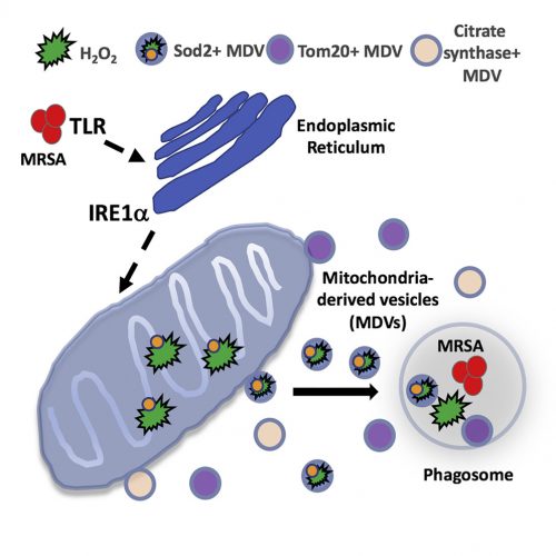
What Abuaita and co-authors describe is a process by which certain bacteria, including the potentially dangerous methicillin-resistant Staphylococcus aureus, trigger more than simply a phagocytic event. Rather, it is clear that the phagosome somehow recognizes the microorganism within its midst, and launches a far more lethal and complex biochemical reaction. The cascade begins with the infected macrophage mounting a stress response – one initiated by the phagosome but further elaborated by the endoplasmic reticulum. Specifically, the endoplasmic reticulum turns on one of the most elegantly choreographed and well-described quality-control systems in the cell, called the unfolded protein response, or UPR. The critical sensor that needs to be tripped to turn on the pathway is the endoplasmic reticulum membrane protein, IRE1a. It is at this point that the cell biology really gets exciting.

The response is not confined to the endoplasmic reticulum and well understood downstream nuclear effector pathways; rather, the signal is somehow transmitted to mitochondria. In response, the organelle is prompted to produce reactive oxygen species – hydrogen peroxide in particular. Mitochondria are known to produce reactive oxygen species, including hydrogen peroxide, as part of their normal energy-generating metabolism. What is different here is that a defense mechanism initiated in the phagosome, signals the endoplasmic reticulum, which then communicates to the mitochondria; a truly elegant and previously unrecognized relay system.

As interesting as the new interorganellar signaling events are – there is more. Specifically, the fascinatingly novel trafficking pathways identified.

The newly synthesized mitochondrial hydrogen peroxide is packaged in vesicles which are shed from the organelle. The authors even identify a critical component of this release step – the ubiquitin ligase, Parkin. These newly created, hydrogen peroxide-loaded mitochondrial vesicles then migrate through the cell, destined to fuse with pathogen-infected phagosomes – releasing their toxic contents and supplementing the already initiated bacterial killing process. Just in case the newly delivered hydrogen peroxide does not provide a sufficient amount of toxic reactive oxygen, some mitochondrial vesicles actually encapsulate the hydrogen peroxide-synthesizing enzyme, superoxide dismutase-2.

What we have here is the cell marshalling its degradative armamentarium in a manner we never imagined to help fight methicillin-resistant Staphylococcus aureus and other invading pathogens. It seems clear that this is only the tip of the iceberg – interorganelle communication/signaling networks and elaborately coordinated effector apparatuses in cells are certain to exist in ways we can only begin to imagine. In this case, the goal is to fight infection; in others, it may very well be to thwart the effects of aging, to counter disease, or neutralize the effects of mutations, toxins, or other environmental insults. The more we know about cell biology, the more we realize how much we do not know; it is amazing how the field continues to captivate our attention.

SRT – February 2019

[1]B.H. Abuaita et al., Cell Host and Microbe (2018) doi: 10.1016/j.chom.2018.10.005. PMID: 30449314 (Request article via ILL).

The Value of Negative Results

Every investigator hopes the results they obtain support the hypothesis they put forth. However, more often than not, this does not happen – the data acquired do not conform to what was expected. What to do then with the “negative” results?

To be clear, the results we are talking about were obtained through carefully considered, well executed, and appropriately controlled (read: presence of positive and negative controls) experiments. They simply do not extend that which was predicted; in some cases, they may even call into question the underlying – often already published – results that led to the hypothesis guiding the study.

Of course, it may be that the work that was to be extended was never solid in the first place. That is, it is possible that the prevailing view in a field is based on incorrect and/or irreproducible results. Indeed, a number of studies are showing an alarmingly large percentage of high-profile published results are not reproducible [1],[2]. In the field of cancer – the pharmaceutical giants Amgen and Bayer Healthcare were unable to replicate the findings included in a large number of studies published in elite journals. The implications are profound; how can companies which rely on such research published in the scientific literature to define molecular targets and develop therapeutic drugs, do so against a backdrop of irreproducibility?

Source: Baker M. 1,500 scientists lift the lid on reproducibility. Nature; 2016.

Why is this happening? Explanations offered include – among others, pressures to publish, financial considerations, poor statistical analyses, insufficiently detailed protocols/technical complexity, selective reporting, and inadequate reagent authentication. The National Institutes of Health is aware of these problems and is calling on investigators seeking support to include in their proposals detailed descriptions of i. the scientific premise of the proposal; ii. experimental design specifications; iii. how biological variability will be considered; and iv. how biological and chemical reagents will be authenticated[3]. This is in addition to newly required statements regarding evidence that a detailed plan for data analysis is in place.

Although the (negative) results obtained do not confirm or extend other studies, the point here is that they are still very much of value. Other scientists in the field would welcome knowing what was done, and what was observed. The obvious benefit is that it will prevent others from wasting time, energy, and resources on approaches that are not fruitful, and would help focus the field by better defining what are, and what are not reliable results/models.

Despite the inherent value of such results, there is the perception of publication bias – the belief that only positive data is worthy of publication. Non-confirmatory or negative results are often not disseminated, at great cost to the scientific community.

One approach to assuring sufficient promulgation of the conclusions of a study is for appropriate journals to agree, in advance, to publish the results regardless of whether they confirm or refute the underlying hypotheses that initiated the study. This would provide a mechanism for a field to enjoy a wealth of otherwise unreported information about what works, and what does not – in particular investigators’ laboratories. Many clinical trials operate this way, with final results disseminated regardless of the effects seen on patients. A second approach would be to develop journals that will consider negative, confirmatory, and non-confirmatory results, data notes, and virtually any valid scientific or technical finding in a particular field. F1000Research is a journal that adheres to just such guidelines. The idea that all results are embraced is extremely attractive; after all, science is, at its core, simply a search for the truth.

SRT – November 2018

[1] M. Baker, Nature (2016) doi: 10.1038/533452a. PMID: 27225100 (article)
[2] C.G. Begley and L.M. Ellis, Nature (2012) doi: 10.1038/483531a. PMID: 22460880 (article)
[3] National Institutes of Health – New Grant Guidelines; what you need to know. https://grants.nih.gov/reproducibility/documents/grant-guideline.pdf

Senolytic Strategies to Combat Aging

Aging is a multifactorial process – with “hallmarks” [1] including genomic instability and altered gene expression, epigenetic effects, telomere erosion, stem cell fatigue, proteostatic errors, senescence, compromised mitochondrial, lysosomal, and peroxisomal function, and disrupted communication networks, among others. Senescence, in particular, is critical as it is thought to constitute a major decision point for cells. With advancing age, cells accumulate sufficient damage and are stressed to the point that they must make a crucial decision – either transform and become cancerous, or pass into senescence.

The anti-tumor senescent state is one of continued metabolic activity, but no cell division. Unfortunately, accompanying the senescent phenotype is cellular release of assorted chemokines, growth factors, interleukins, and proteases. The ultimate effect of this collection of pro-inflammatory mediators is to corrupt the regional cell/tissue milieu. Ironically, this newly created environment is very conducive to cellular transformation and tumor development.  Indeed, investigators in the field refer to the senescence-associated secretory phenotype as “the dark side of tumor suppression”[2].

What if the decision to pass into senescence could be maintained (and thus avoid cancerous transformation), but the senescent cell could somehow be destroyed? Would cancer be averted and cell/tissue integrity maintained?  Could stem cell exhaustion be ameliorated and health span increased?  Could aging, the ultimate risk factor for human disease, be brought under control – at least to some extent? While clear answers to these questions remain elusive, the idea of trying to destroy senescent cells – either through genetic means or specific drugs (called “senolytics”), is a major focus of researchers in the field of cellular aging.

Aged hand with pill
Do senolytics hold the key to healthy aging?

Dr. Van Deursen’s group at the Mayo Clinic College of Medicine developed a brilliant strategy of induced senescent cell suicide. In mouse models, they created a genetic background whereby cells would die the moment they expressed the senescence biomarker p16Ink4A. They showed both in progeroid[3] (i.e., prematurely aged) and wild-type[4] backgrounds, that targeted elimination of senescent cells improved the health of the animals by delaying, or preventing, impaired tissue function. Furthermore, in the wild-type background, the mice lived some 30% longer!

Instead of genetic approaches, investigators from Dr. Robbins’ group at the Scripps Research Institute screened for drugs that would selectively target senescent cells – while not affecting other cells. Interestingly, inhibitors of HSP90 were identified [5] as potent senolytics – with efficacy in both in vitro (i.e., cellular) and in vivo (i.e., whole animal) models. HSP90s are critical cellular chaperones, which facilitate protein folding and stabilize hundreds of molecules involved in a myriad of biochemical and metabolic pathways. The paper’s authors speculate that inhibiting HSP90’s anti-apoptotic (i.e., cell death)/pro-survival activities may be playing some role in selectively targeting senescent cells – a view consistent with the activity of other demonstrated senolytics[6] that are thought to reduce resistance to apoptosis.

How to partner senolytics with other drugs to maximize efficient elimination of senescent cells while limiting toxicity is an important focus going forward. Although the science is not quite there yet – the notion of clinical trials employing senoltyics or other modulators of cell senescence with the goal of thwarting aging’s effects and improving health span are not that far off.


SRT – September 2018

[1]C. Lopez-Otin et al., Cell. (2013) doi: 10.1016/j.cell.2013.05.039. (PMID: 23746838)
[2] J-P. Coppe et al., Ann Rev Pathol. (2010) doi: 10.1146/annurev-pathol-121808-102144 (PMID:20078217)
[3] D.J. Baker et al., Nature (2011) doi: 10.1038/nature10600. (PMID: 22048312)
[4] D.J. Baker et al., Nature (2016) doi: 10.1038/nature16932. (PMID: 26840489)
[5] H. Fuhrmann-Stroissnigg et al., Nat Commun. (2017) doi: 10.1038/s41467-017-00314-z. (PMID: 28871086)
[6]A. Hernandez-Segura et al., Trends in Cell Biology (2018) doi: 10.1016/j.tcb.2018.02.001 (PMID: 29477613)

CRISPR-Cas9 and p53

The CRISPR-Cas9 system represents a remarkable breakthrough in genome editing technology. With relative ease and amazing precision, investigators may now alter or replace genes in the genomes of organisms across the evolutionary spectrum. The potential for human disease treatment or prevention is incredible – an observation not lost on the pharmaceutical companies that have invested heavily in the approach.

All the excitement notwithstanding, some problems have arisen with the technology that may limit its usefulness going forward. Specifically, two papers from the journal Nature Medicine[1],[2] suggest that the “genome guardian” protein, p53, is activated in response to employment of CRISPR-Cas9. To understand what p53 is responding to, a brief description of how the CRISPR-Cas 9 system works is warranted.

The CRISPR (clustered regularly interspaced short palindromic repeats)-Cas9 (CRISPR associated protein 9) system employs specific enzymes and carefully designed RNA guides to seek out distinct genomic DNA sequences for editing, removal, or replacement. The process involves the cleavage of DNA – an action not unnoticed by the human cell. Indeed, it is at this step that the cell takes exception to indiscriminate cutting of its DNA and turns on the (DNA) damage response pathway – orchestrated by p53. The aforementioned papers document the effects in human cells of such initiation of the p53-mediated reaction to DNA strand breaks.

CRISPR Cas9 system (Source: Marius Walter; Wikimedia Commons)

In the paper by Haapaniemi and colleagues, the authors observe that CRISPR-Cas9 activates the p53 pathway in human cells, and limits the efficiency of the gene editing process. In contrast, inhibiting the damage response pathway allows for greater effectiveness of the CRISPR-Cas9 genomic modification apparatus. The obvious problem is that any attempts to down-regulate p53, or to select for cells that have reduced activity of the pathway, by definition create cells potentially susceptible to cancerous transformation. The authors suggest that in future work, mechanisms by which the DNA repair pathway could be modulated – perhaps turned off during gene editing, but reinitiated shortly thereafter, represent an appropriate work around.

In the study by Ihry and colleagues, attempts are made to employ CRISPR-Cas9 to reprogram pluripotent stem cells. Once again, the gene editing procedure elicits a p53-mediated DNA damage response, and the cells undergo programmed cell death (~apoptosis). Cells that survive are very much at risk as they may harbor down-regulated p53 – potentiating the possibility of an accumulation of offsite mutations. As above, the answer seems to point to modulating p53 activity – turning it off for the editing step, but reactivating immediately following.

Much has been written about these results, both in the scientific literature and popular press. No one is calling for the technique to be abandoned; rather, additional research will be required before it can be expected that the human cell’s sophisticated and evolutionarily developed capacity for protecting itself can be sidestepped. Expect a wealth of research to be published on the topic in coming months. Stay tuned!

SRT – August 2018

References:
[1] E. Haapaniemi et al., Nat Med. (2018) doi: 10.1038/s41591-018-0049-z. [Epub ahead of print] (Link to Text)

[2] R.J. Ihry et al., Nat Med. (2018) doi: 10.1038/s41591-018-0050-6. Epub 2018 Jun 11. (Link to Text).

Targeted Approach to Antimalarial Drug Development

Hello all and welcome to this corner of the Interprofessional Health Sciences Library and Information Commons website, an area devoted to bringing you what we hope you find interesting and exciting stories of innovation in biomedical science.  Thank you for joining us!

A photomicrograph of a blood smear that contains a macro- and microgametocyte of the Plasmodium falciparum parasite. Source: CDC/Dr. Mae Melvin Transwiki

This first installment focuses on malaria – a worldwide health problem that, according to the Centers for Disease Control and Prevention, killed some 445,000 people in 2016.  Malaria is caused by parasites, carried by mosquitos which introduce the microorganisms into humans’ blood. Infection results in flu-like fever/chills, headache, and respiratory problems. Left untreated – progression to more serious and life-threatening complications is rapid. The parasite most responsible for this scourge is the protozoan Plasmodium falciparum. Pharmacological interventions exist – but have been thwarted by the unicellular organisms’ ability to acquire resistance. For example, chloroquine and related compounds were employed for decades to disable the parasite’s lysosome-like digestive vacuole. With the organism unable to completely metabolize the host red blood cells’ hemoglobin, toxic metabolites amass and the parasites die. The more recent drug of choice is artemisinin, a natural product derived from the plant Artemisia annua. Compounds containing artemisinin, or related semi-synthetic derivatives, reduce the number of parasites in the bloodstream through what is thought to involve formation of highly reactive free radical species. Unfortunately, identification of other safe drugs with efficacy in killing the microorganism has not been successful.

Which brings us to the new approach – pioneered by a team of investigators of the University of South Florida and Wellcome Trust Sanger Institute and described in a research article entitled “Uncovering the essential genes of the human malarial parasite Plasmodium falciparum by saturation mutagenesis”[1].  Here, a technique called piggyback transposition mutagenesis was used to insert crippling DNA stretches into genes across the organism’s genome. Non-essential genes were so-identified by their ability to withstand the random insertions. Essential genes, in contrast, resulted in the parasite’s death. Illumina-based deep sequencing allowed for identification of the insertion sites – and the analysis was on!

Of the 5399 genes that exist in the P. falciparum genome, 2680 were found to be essential for the parasite’s disease propagation processes. Powerful bioinformatic analyses revealed that essential gene clusters exist encoding proteins involved in, among other functions – lipid metabolic pathways, proteasomal degradation, cell cycle control, and RNA stability.  Naturally, these genes and their associated proteins/pathways now are the molecular targets for future antimalarial drug development. It is certain that sophisticated drug screens are currently being employed and we can only hope that safe and efficacious compounds are found in a timely manner. With the dearth of currently available antimalarials and the disease’s continued health toll, advances cannot come too soon.

SRT – June 2018

Reference:
[1] M. Zhang et al., Science 360, eaap7847 (2018). DOI: 10:1126/science.aap7847 (Link to Text)