Call for Submissions: COVID-19 Oral History Project

COVID-19 oral history call for submissionsTo reconnect as a community during the COVID-19 pandemic, SHU University Libraries seek your stories of what this time has been like for you. We hope that sharing these stories with one another will bring us back together in a new way, through sharing our personal experiences of this moment.

HOW TO PARTICIPATE:

Please record a 1-3 minute narrative about your experience, using any video or audio equipment available to you, and submit the file to our e-Repository (Under “Author Corner”: Submit Contribution in the left column. If you don’t have an account, you will need to create one).  Please also submit an image that represents your narrative, which will appear next to your recording in the published archive.

Questions to guide your response:

  • What is your day to day life like? What would you want people the future to know about what life is like for us now?
  • What has been most challenging about this time? What do you miss about your life before COVID-19? Are there specific places or things on campus that you miss?
  • Essential is a word we are hearing a lot right now. What does essential mean to you? Who is essential? What are we learning about what is essential?
  • What is COVID-19 making possible that never existed before? What good do you see coming out of this moment? How can we re-frame this moment as an opportunity?
  • What is it you want to remember about this time? What have you learned?
  • After this pandemic ends, will things go back to the way they were? What kinds of changes would you like to see? How will you contribute to rebuilding the world? What will you do differently?

With thanks to the scholars and librarians who came together to create this project: Professors Angela Kariotis Kotsonis, Sharon Ince, Marta Deyrup, Lisa DeLuca, and Alan Delozier, Technical Services Archivist Sheridan Sayles and Assistant Deans Elizabeth Leonard and Sarah Ponichtera.

Information Resources on Coronavirus Disease 2019 (COVID-19)

Update (2.28.2020): We’ve migrated the below information to the Information Resources on Coronavirus Disease 2019 (COVID-19) toolkit. This toolkit will be updated as additional resources and information is published.

The IHS Library recommends consulting the following resources for factual, up-to-date information on Coronavirus Disease 2019 (COVID-19).

Health Agency Information:

Coronavirus Disease 2019 (Centers for Disease Control and Prevention): Contains information about what you should know about the Coronavirus, situation updates, including a list of locations of confirmed cases, and information resources for travelers and healthcare professionals.

Coronavirus disease (COVID-19) outbreak (World Health Organization): Contains rolling updates about the disease outbreak, as well as important myth-busters to stop the spread of false information. You may also wish to take the WHO e-learning module, Emerging respiratory viruses, including COVID-19: methods for detection, prevention, response and control.

Research Resources:

2019-nCoV (PubMed): A preformulated search will bring up the latest research from the National Library of Medicine’s PubMed Database.

Coronavirus Disease 2019 (JAMA): JAMA Network’s updates on coronavirus diagnosis and treatment, along with recent articles.

Novel Coronavirus Information Center (Elsevier): Elsevier is providing free health and medical research on COVID-19, which includes access to the Coronavirus Research Repository.

Information for Patients:

Coronavirus Infections (MedlinePlus): MedlinePlus is a service of the National Library of Medicine (NLM), which provides quality, plain-language information for patients.

New Artwork Unveiled at the IHS Library

Neuron art print
“Neuron” by Andrea P. Tóth.

Earlier this month, twelve colorful anatomy prints were permanently installed in various locations throughout the library. The mixed-media watercolor and ink designs are by Prague artist and medical doctor, Andrea P. Tóth, owner of the small-business MedPapers.

When the IHS Library opened in July 2018, the space was beautiful- but the white walls were noticeably in need of artwork. Looking for help on this huge project, the IHS Library consulted with the Hackensack Meridian School of Medicine’s Art and Medicine student group for design inspiration. Toth’s prints were ultimately selected for the library for their color, creative interpretation of the human body, and tone of calmness.

So far, the prints have been a huge hit with everyone, especially IHS Library Director, Chris Duffy. “We couldn’t be happier with how these beautiful prints look in our library,” he says. “I hope you enjoy them as much as we do!”

Information Commons
Six prints adorn one wall of the Information Commons.
Neuron synapse art
“Neuron Synapse” by Andrea P. Tóth.

Caenorhabditis elegans – the Quintessential Biological Model

The nematode Caenorhabditis elegans (C. elegans) has proven itself time and time again to be an organism of immense value to biomedical researchers. Important studies employing the biological model appear on a regular basis in top tier journals across a wide array of research areas. One perusing the scientific literature is reminded of the worm’s immense power quite regularly. Consider, for example, two related papers that appeared recently in the press; one regarding selective autophagy and lifespan[1], and the other focused on caloric restriction and how its anti-aging effects are elicited from a cellular/metabolic perspective[2].

C. elegans 3D model
The C. elegans 3D model. VirtualWorm project.

In the former, Kumsta and colleagues show that the (C. elegans) protein p62/SQST-1 (~p62) plays an important role in recognizing cellular proteins, macromolecular structures, and even intracellular organelles – e.g., mitochondria, earmarked for destruction. Such recognition leads to trafficking of the p62-substrate to intracellular degradative centers where the actual destruction takes place. Importantly, worms genetically engineered to overexpress p62 enjoy not only an efficiently operating “selective autophagy” pathway, but also a 25% increase in lifespan. Which proteins precisely constitute the repertoire of those recognized by p62 remains to be determined, but the idea that selective autophagy is an extant mechanism in cells suggests myriad potential applications in targeting for destruction those proteins or structures identified as toxic, and associated with human disease. Cellular quality control is critical – and surveillance systems including those mediated by p62 that help maintain proteostasis (i.e., integrity of cell proteome) are essential.

The article by Weir and colleagues suggests that the anti-aging effects of caloric restriction are elicited, at least in part, through maintenance of mitochondrial network integrity, and an interplay with functional (i.e., fatty acid metabolizing) peroxisomes. AMP-activated protein kinase (AMPK) acts similarly to dietary restriction, eliciting many equivalent effects – including those on longevity. These studies beg the follow up question – are anti-aging therapeutics of the future those that both assure structurally sound mitochondria whose metabolic (read: fat metabolizing) functions are carefully coordinated with peroxisomes, and activate appropriate metabolic cascades – including those involving AMPK? The data generated with C. elegans and presented in this interesting (Cell Metabolism) paper certainly supports such conclusions.


A final word or two regarding C. elegans. Dr. Sydney Brenner performed pioneering work in the 1960s and 1970s establishing the organism as a powerful model for biomedical studies. Among the work done was a description of the worm’s neuronal circuitry. For these and related studies, Dr. Brenner, and Drs. H. Robert Horvitz and John Sulston were awarded the 2002 Nobel Prize in Physiology and Medicine. C. elegans was the first multicellular eukaryote to have its genome sequenced; the developmental outcome of every one of its 959 of its cells is known; and all its neural connections are identified. The latter, known as a “connectome”, is available in no other animal at present. The worm has been used in studies involving myriad topics in cell biology, with results impacting all aspects of human health, disease, and aging.

The organism made big news when it was revealed in 2003 that nematodes brought aboard the shuttle Columbia for experimental purposes, had survived the tragic fiery crash of the spacecraft. Upon reentry into the earth’s atmosphere, the creatures were exposed to astonishingly harsh temperatures, centrifugal/gravitational forces, and atmospheric conditions; yet they returned alive. If worms could survive such conditions – could other microorganisms also do so?  Over the course of time, have microorganisms hitched rides on asteroids, comets, meteors and the like and traveled across the heavens – transferring life forms? Hmm…

SRT – January 2020

References:

[1] Kumsta C, Chang JT, Lee R, et al. The autophagy receptor p62/SQST-1 promotes proteostasis and longevity in C. elegans by inducing autophagy. Nat Commun. 2019;10(1):5648. Published 2019 Dec 11. doi:10.1038/s41467-019-13540-4

[2] Weir HJ, Yao P, Huynh FK, et al. Dietary Restriction and AMPK Increase Lifespan via Mitochondrial Network and Peroxisome Remodeling. Cell Metab. 2017;26(6):884–896.e5. doi:10.1016/j.cmet.2017.09.024

APA 7th Edition: What’s new?

APA 7th EditionBy Kyle Downey, Health Sciences Librarian

Back in October the American Psychological Association (APA) released the 7th edition of their APA Publication Manual.  It has been nearly a decade since the 6th edition was released and with this newest edition we see several additions and revisions.

So, what is new?

Some changes to the new publication will be immediately noticeable to the user who has used the previous 6th edition.  First, the new manual is in full color throughout the entire publication.  Some other changes include:

  • Citing of online material, with a focus on social media
  • Inclusion of bias-free language
  • Guidelines on writing without bias that addresses age, disability, gender, race and ethnicity, including the singular use of “they”
  • Using shortened URLs and shortDOIs if a URL or DOI is long and complex
  • Removal of publisher locations for books and book chapters
  • An in-text citation with 3 or more authors is to be shortened to include only the first authors name and “et al”
  • Website URLs no longer need to be preceded with “Retrieved from” unless there is also a retrieval date
  • A single space after any body-text punctuation rather than 2 spaces

To learn more about the new publication manual, check out the APA style blog.

Both the Walsh Library and the IHS Library also have permanent reference copies available for faculty and students to use.

Source: Elias, Daniel. “APA Style 7th Edition: What’s Changed?” MyBib, MyBib, 14 Sept. 2019.

Get articles easier with Libkey Nomad Chrome extension!

We are excited to announce a new tool that will enhance and simplify the research experience for our IHS community: Libkey Nomad.

Libkey Nomad, a browser extension for Google Chrome, automatically links to full-text content from websites such as PubMed, Wikipedia, and Google Scholar.  To quote Third Iron, “Libkey Nomad keeps libraries at the heart of the research process by connecting researchers to library content, even when the user is not in the library.”

Installation instructions for Libkey Nomad can be found here.

If you have questions or need help with downloading the extension, contact you IHS librarian directly or via ihslibrary@shu.edu.

Libkey Nomad Chrome extension

A Bioethicist’s Response to “When is a Brain Really Dead?”

Today’s post is a response by Dr. Bryan Pilkington to Dr. Stan Terlecky’s July 15, 2019 Terlecky’s Corner post entitled, “When is a Brain Really Dead?” Dr. Pilkington is an Associate Professor at Seton Hall University.

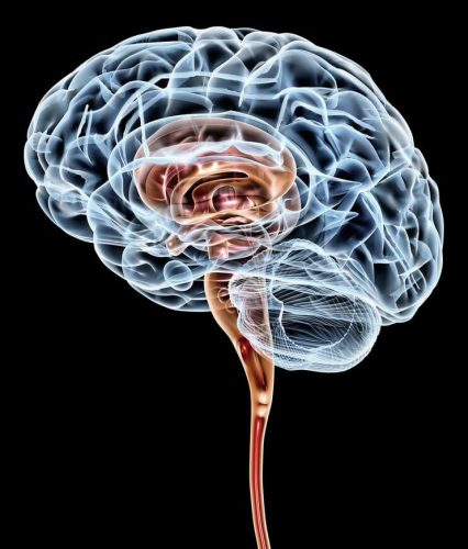
Last month’s Terlecky’s Corner post raises the question, “When is a brain really dead?” The reanimation – to borrow Terlecky’s phrase and his caution about its use – of porcine brains is both exciting and concerning. The excitement is due, as Terlecky notes well, to the possible benefits of such work: “potentially impacting several health scourges of our time including Alzheimer’s disease, Parkinson’s disease, and other age-related neurodegenerative disorders.”[1] He also notices that ethicists will be busy sorting through these and future studies, especially if the human brain gets involved.

Brain

This is how the usual dialectic goes when addressing ethical questions about emerging medical technologies. Undoubtedly, there will be lectures and papers from ethicists raising the usual kinds of questions, many titled with some version of, “We can, but should we?” These are not endeavors without merit; ethical analysis often lags behind research; in many cases, something has to be created in order to have something about which to raise questions. In fact, one of the important tasks of ethicists in this realm is to serve as watchdogs: to give caution, to aid researchers in thinking through possible concerns, and in some instances – at least with respect to IRB oversight – to say “no.”[2] However, though this is a useful and important role, it comes at a cost. Many times, those raising these kinds of concerns are termed “the ethics police”[3] and this image has hurt collaboration on research and made more challenging the necessary interprofessional tasks that medical research and healthcare practices are.

So what should be said in response to this kind of research? What are the answers to Terlecky’s list of ethical questions? The questions require longer answers than the space of a blog post admits, but I’ll take a shot at two sets of questions, suggesting a model for answering them and those remaining.

The first set comprises a list of real and immediate questions about one of the practical consequences of this research: organ donation. Will donation rates plummet? Will the organ shortage increase because there will be hold outs who think reanimation is possible? Will public trust diminish as this research moves forward?[4] Will families request that additional healthcare resources be spent on loved ones who meet the legal definition of brain death? An answer which avoids the problematic policing approach but takes seriously the importance of these questions is to engage in an extended conversation with researchers, ethicists, healthcare practitioners and administrators, and members of their communities about new technology, how it might be used, and its far-reaching implications. We must not shy away from these hard questions nor from recognizing the potential value of certain kinds of research, but we must also keep in mind the possible negative externalities that could result. This approach raises more questions. Should this research be halted if it damages public trust? Should it be stopped if fewer organs are donated? These questions lead to further questions. If the organ shortage is a primary concern, is it appropriate to connect it to this research? The conversation I am suggesting must consider that, as well. Some have argued that the sale of organs should be allowed and that this would alleviate the shortage[5], others have raised concerns about the commodification of human beings if such sale is legalized[6] – the breadth of the needed conversation is wide, as answers to questions about organ sale are connected to ethical concerns raised about porcine brain experimentation.

A second set of questions hover around the definition of death and the sources upon which we rely to answer those questions. Terlecky helpfully asks about the legal, ethical, and spiritual determinations of death. From where or in what do we root our conceptions of life, of human flourishing, and of death? Are they religious? Are they legalistic? Are they rooted in metaphysical conceptions of the person that we learned in our undergraduate philosophy courses? The kind of conversation I am suggesting is most effectively held when we bring the deep and rich traditions that inform our thought to bear on the subject matter under discussion. It is not a simple task to work through various traditions and try to understand how others think about reanimated porcine brains, human brains, and death, but that is what is needed. As physician and ethicist Lauris Kaldjian recently asked of his colleagues,[7] did you take a course about the existential questions in medical school? Though rhetorical, the suggestion is powerful. How should we respond to this research? In the same way we should respond to all research that raises significant ethical questions, by practically reasoning together.


References:

[1] Terlecky, S. 2019, July 15. “When is a Brain Really Dead?

[2] ​Evans, J. 2012. The History and Future of Bioethics: A Sociological Account. Oxford, United​ ​Kingdom: Oxford University Press.

[3] Klitzman, R. 2015. The Ethics Police? The Struggle to Make Human Research Safe. Oxford: Oxford University Press

[4] ​Moschella, M. 2018. Brain death and organ donation: A crisis of public trust. Christian Bioethics 24(2):133–50.

[5] ​Cherry, M. ​2005​. ​K​​idney for Sale by Owner: Human Organs, Transplantation, and the Market. Georgetown University Press.

[6] Pilkington, B. 2018. A Market in Human Flesh: Ramsey’s Argument on Organ Sale, 50 years later. Christian Bioethics 24(2):133–50.

[7] Kaldjian, L. (Personal communication during lecture in Grand Rapids, Michigan, March 25, 2019).

When is a Brain Really Dead?

When is a brain really dead? The answer to this question was made far more complicated by the recent work of Dr. Nenad Sestan and colleagues at Yale University. Their astonishing paper entitled “Restoration of brain circulation and cellular functions post-mortem,” appeared in the journal Nature[1] this April. In it, the authors were able to demonstrate that brains taken from slaughtered animals (pigs in this case), could be “reanimated” 4 hours later in the laboratory – and made at least partially functional for some 6 hours thereafter. I use the word reanimated with some trepidation – it implies the brains were dead and somehow brought back to life. That is not quite the story – rather, the organ turns out to be far more resilient than we previously realized and the research team simply identified a way to tap into that inherent resiliency.

A brief description of the research study: 32 brains from slaughtered pigs were delivered to the research team on ice. Within 4 hours, the scientists carefully perfused the brains using a proprietary surgical procedure, pumping apparatus, and oxygen and nutrient-rich solution collectively termed BrainEx. The team then analyzed the brains for specific cellular, metabolic, and electrical activities. What they found was incredible. The BrainEx perfusion system restored a number of brain functions, including glucose and O2 utilization and concomitant CO2 production (indicative of metabolic function), induced inflammatory responses (suggesting an active immune system), active microcirculation (evidence of structural integrity), and electrical activity (with neuronal firing).

Figure of porcine brain connected to perfusion system
Connection of the porcine brain to the perfusion system via arterial lines. The pulse generator (PG) transforms continuous flow to pulsatile perfusion. Source: Figure 1B, Nature 568, 336–343 (2019).

With respect to the last point, it should be noted the investigators were well aware of the ethical concern that full restoration of brain function could potentially lead to a state of “consciousness.” What it would mean for a disembodied brain to attempt to operate without peripheral sensory input, and what the organ might remember, was simply too much to consider; the brains were treated pharmacologically to assure no coordinated higher level cognitive activity was possible. Said another way, the renewed brains could not begin to think.

Overall, the results suggested to a first approximation, functional activity of the otherwise dead brain had been restored. Importantly from an experimental standpoint, control perfusates were without effect – the brains degenerated much like untreated specimens.

Against the backdrop of these remarkable results are the inevitable ethical questions that follow. Returning to the one posed above – when is a brain really dead? If our understanding of brain death requires a deeper examination, how then do we determine legally, ethically, and/or spiritually, when a person truly dies? What are the implications of this work on the issue of organ donations? Is a brain death as currently defined sufficient to permit the harvesting of a person’s organs? Will (and should) donations ebb as people begin to question the legitimacy of declarations of death. Also, could future brain reanimation experiments include restoration of conscious thought? Certainly the ability to control the brain in this manner permits the testing of drugs in new ways – potentially impacting several health scourges of our time including Alzheimer’s disease, Parkinson’s disease, and other age-related neurodegenerative disorders.

Naturally, science requires replication, expansion, and more careful delineation of what is, and is not possible with the technology described. But what enormous doors have been opened for neuroscientists, neurologists, neuropharmacologists, cognitive scientists, and the many others interested in the structure and function of the brain. I suspect ethicists will also be very busy sorting through these and the inevitable follow-up studies – especially as applications involving the human brain are contemplated.

SRT – July 2019

[1] Vrselja Z, et al., Nature 568, 336-343 (2019). PMID: 30996318

Cardiac Exosomes to the Rescue

In an extremely exciting study that appeared last month in the journal Nature Communications[1], scientists from the University of Alabama at Birmingham, and Huazhong University of Science and Technology in Wuhan, China, showed that after myocardial infarction (~heart attack), heart tissue release tiny membrane-bound vesicles called exosomes, specifically loaded with genetic material designed to promote cardiac tissue repair. Such restoration is mediated by bone marrow progenitor cells that have been released from their sequestered bone-specific existence by the information carried in the newly arrived exosomes.

Several hundred thousand Americans suffer heart attacks each year, making it critical for the medical and scientific communities to understand this potentially transformative study. To begin our analysis, let’s begin with exosomes – tiny membrane-enclosed vesicles released from many cell types. Thought to mediate communication/cell-cell signaling activities, exosomes leave cells loaded with proteins, lipids, DNA, mRNA, and microRNAs among other potentially bioactive molecules. Exosomes are synthesized within the endosome-lysosome system of cells – emerging cargo loaded and poised to deliver their contents locally, or at a distance. It is perhaps not surprising that several pharmaceutical companies – recognizing the potential drug delivery platform that exosomes represent, are pursuing the tiny vesicles in several biomedical contexts.

Once released, how the nascent exosomes hone to specific target tissues and cells is unclear. Perhaps the proteins assembled into their membranes – distinct to each type of exosome – play a role in this discrimination. Also not well understood is the nature of the exosome-target cell interaction. Is it a receptor-ligand-like binding event that triggers signaling events and results in fusion and integration of exosomal contents? Or is it a more basic fusion of two membranes held in close apposition? What about a straightforward endocytic process whereby the exosome is taken up and released within the newly formed endosome?

Back to the study at hand – the investigative team identified the exosomes released from damaged heart muscle as containing specific microRNAs (called myocardial microRNAs or myo-miRs). Recall microRNAs are short, non-coding RNAs with the capacity to (negatively) regulate gene expression. The myo-miRs under consideration here do just that – they very effectively block activity of the chemokine receptor, CXCR4 in bone marrow progenitor cells. Such inhibition allows such stem-like cells to escape their seclusion, enter the bloodstream, and travel to the injured heart…“exosomes to the rescue.” There, repair processes are initiated.

Knowing this cell biology, there must be ways to better harness exosomes to improve cardiac repair. Investigators are sure to pursue this quickly. Almost as assuredly, scientists will also be looking for other examples of exosomes and their bioactive contents as biomarkers for pathology, and as potential rescue mechanisms for disease’s damaging wrath.

SRT – April 2019

[1]Cheng M, et al., Nat Commun. (2019) doi: 10.1038/s41467-019-08895-

My Professional Journey: Dr. Heather Frimmer

On March 13, Hackensack Meridian School of Medicine at SHU students, faculty, and staff gathered over lunch to hear Dr. Heather Frimmer discuss her unique career trajectory from medical trainee to radiologist and published author. This engaging event was part of the “My Professional Journey” series sponsored by Student Affairs and Wellbeing “Careers in Medicine,” the OME Professionalism Committee, and the IHS Library.

During the 90-minute program, Dr. Frimmer spoke of the path that led to her current position as Diagnostic Radiologist in Breast and Emergency Radiology. Once established in her medical career, she would then follow an altogether different path and write her first novel, Bedside Manners, published by SparkPress in 2018.

After her talk, those in attendance were treated to a reading of two passages from Bedside Manners, followed by small-group discussion and reflection of the passages’ themes. Students were then invited to do a bit of their own creative writing based on their personal clinical experiences thus far.

On behalf of everyone who attended, the sponsors of the “My Professional Journey” series would like to thank Dr. Frimmer for joining us and speaking about her medical and writing career.

Dr. Heather Frimmer (left) with former medical school classmate, Dr. Miriam Hoffman, Associate Dean of Medical Education.
From left to right: Dr. Miriam Hoffman (Associate Dean of Medical Education), Janae Moment (MS1), Raquel Cancho Otero (MS1), Dr. Heather Frimmer, Candace Pallitto (MS1), Caryn Katz Loffman (Human Dimension Assistant Course Director), Katherine Veltri (MS1), and Allison Piazza (Health Sciences Librarian).
Medical students and faculty discussing Bedside Manners by Dr. Heather Frimmer.