When is a Brain Really Dead?

When is a brain really dead? The answer to this question was made far more complicated by the recent work of Dr. Nenad Sestan and colleagues at Yale University. Their astonishing paper entitled “Restoration of brain circulation and cellular functions post-mortem,” appeared in the journal Nature[1] this April. In it, the authors were able to demonstrate that brains taken from slaughtered animals (pigs in this case), could be “reanimated” 4 hours later in the laboratory – and made at least partially functional for some 6 hours thereafter. I use the word reanimated with some trepidation – it implies the brains were dead and somehow brought back to life. That is not quite the story – rather, the organ turns out to be far more resilient than we previously realized and the research team simply identified a way to tap into that inherent resiliency.

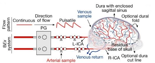
A brief description of the research study: 32 brains from slaughtered pigs were delivered to the research team on ice. Within 4 hours, the scientists carefully perfused the brains using a proprietary surgical procedure, pumping apparatus, and oxygen and nutrient-rich solution collectively termed BrainEx. The team then analyzed the brains for specific cellular, metabolic, and electrical activities. What they found was incredible. The BrainEx perfusion system restored a number of brain functions, including glucose and O2 utilization and concomitant CO2 production (indicative of metabolic function), induced inflammatory responses (suggesting an active immune system), active microcirculation (evidence of structural integrity), and electrical activity (with neuronal firing).

Figure of porcine brain connected to perfusion system
Connection of the porcine brain to the perfusion system via arterial lines. The pulse generator (PG) transforms continuous flow to pulsatile perfusion. Source: Figure 1B, Nature 568, 336–343 (2019).

With respect to the last point, it should be noted the investigators were well aware of the ethical concern that full restoration of brain function could potentially lead to a state of “consciousness.” What it would mean for a disembodied brain to attempt to operate without peripheral sensory input, and what the organ might remember, was simply too much to consider; the brains were treated pharmacologically to assure no coordinated higher level cognitive activity was possible. Said another way, the renewed brains could not begin to think.

Overall, the results suggested to a first approximation, functional activity of the otherwise dead brain had been restored. Importantly from an experimental standpoint, control perfusates were without effect – the brains degenerated much like untreated specimens.

Against the backdrop of these remarkable results are the inevitable ethical questions that follow. Returning to the one posed above – when is a brain really dead? If our understanding of brain death requires a deeper examination, how then do we determine legally, ethically, and/or spiritually, when a person truly dies? What are the implications of this work on the issue of organ donations? Is a brain death as currently defined sufficient to permit the harvesting of a person’s organs? Will (and should) donations ebb as people begin to question the legitimacy of declarations of death. Also, could future brain reanimation experiments include restoration of conscious thought? Certainly the ability to control the brain in this manner permits the testing of drugs in new ways – potentially impacting several health scourges of our time including Alzheimer’s disease, Parkinson’s disease, and other age-related neurodegenerative disorders.

Naturally, science requires replication, expansion, and more careful delineation of what is, and is not possible with the technology described. But what enormous doors have been opened for neuroscientists, neurologists, neuropharmacologists, cognitive scientists, and the many others interested in the structure and function of the brain. I suspect ethicists will also be very busy sorting through these and the inevitable follow-up studies – especially as applications involving the human brain are contemplated.

SRT – July 2019

[1] Vrselja Z, et al., Nature 568, 336-343 (2019). PMID: 30996318

Faster and easier article PDFs in PubMed

We’re pleased to announce another enhancement to the user experience for IHS students, faculty, and staff: Libkey Link for PubMed. To quote Third Iron, the vendor we’ve partnered with for this feature, Libkey Link “brings one-click access to PDF articles in PubMed, dramatically simplifying workflow and improving user experience. No more confusion over what full text source to pick, no more waiting for different pages to load hunting for the PDF button.” We’ve implemented LibKey Link to cut out a couple of steps in the old process, making it easier and faster for you to get the article PDF you’re looking for. As always, if you need any help with this feature (or anything else information-related), contact your IHS librarian.

Improvements to the SHUhealth search tool

SHU Health is a tool that allows IHS Library users to search both the catalog (books, videos, CDs, etc.) and articles simultaneously. We are excited to announce a number of improvements to the interface to streamline the experience for you.

You can search SHUhealth from the library homepage
Getting to SHUhealth from the library homepage

The updated interface is significantly simpler, reducing clutter by removing little-used and unnecessary features. IHS librarians reached out to our partner EBSCO, which manages the interface, to suggest the changes.

Updated and simplified SHUhealth interface
The updated and streamlined SHUhealth interface

We’re interested in continually improving your experience with IHS Library website and other library interfaces. If you have an improvement to suggest or notice a bug we need to fix, email me at andrew.hickner AT shu.edu.

Cardiac Exosomes to the Rescue

In an extremely exciting study that appeared last month in the journal Nature Communications[1], scientists from the University of Alabama at Birmingham, and Huazhong University of Science and Technology in Wuhan, China, showed that after myocardial infarction (~heart attack), heart tissue release tiny membrane-bound vesicles called exosomes, specifically loaded with genetic material designed to promote cardiac tissue repair. Such restoration is mediated by bone marrow progenitor cells that have been released from their sequestered bone-specific existence by the information carried in the newly arrived exosomes.

Several hundred thousand Americans suffer heart attacks each year, making it critical for the medical and scientific communities to understand this potentially transformative study. To begin our analysis, let’s begin with exosomes – tiny membrane-enclosed vesicles released from many cell types. Thought to mediate communication/cell-cell signaling activities, exosomes leave cells loaded with proteins, lipids, DNA, mRNA, and microRNAs among other potentially bioactive molecules. Exosomes are synthesized within the endosome-lysosome system of cells – emerging cargo loaded and poised to deliver their contents locally, or at a distance. It is perhaps not surprising that several pharmaceutical companies – recognizing the potential drug delivery platform that exosomes represent, are pursuing the tiny vesicles in several biomedical contexts.

Once released, how the nascent exosomes hone to specific target tissues and cells is unclear. Perhaps the proteins assembled into their membranes – distinct to each type of exosome – play a role in this discrimination. Also not well understood is the nature of the exosome-target cell interaction. Is it a receptor-ligand-like binding event that triggers signaling events and results in fusion and integration of exosomal contents? Or is it a more basic fusion of two membranes held in close apposition? What about a straightforward endocytic process whereby the exosome is taken up and released within the newly formed endosome?

Back to the study at hand – the investigative team identified the exosomes released from damaged heart muscle as containing specific microRNAs (called myocardial microRNAs or myo-miRs). Recall microRNAs are short, non-coding RNAs with the capacity to (negatively) regulate gene expression. The myo-miRs under consideration here do just that – they very effectively block activity of the chemokine receptor, CXCR4 in bone marrow progenitor cells. Such inhibition allows such stem-like cells to escape their seclusion, enter the bloodstream, and travel to the injured heart…“exosomes to the rescue.” There, repair processes are initiated.

Knowing this cell biology, there must be ways to better harness exosomes to improve cardiac repair. Investigators are sure to pursue this quickly. Almost as assuredly, scientists will also be looking for other examples of exosomes and their bioactive contents as biomarkers for pathology, and as potential rescue mechanisms for disease’s damaging wrath.

SRT – April 2019

[1]Cheng M, et al., Nat Commun. (2019) doi: 10.1038/s41467-019-08895-

My Professional Journey: Dr. Heather Frimmer

On March 13, Hackensack Meridian School of Medicine at SHU students, faculty, and staff gathered over lunch to hear Dr. Heather Frimmer discuss her unique career trajectory from medical trainee to radiologist and published author. This engaging event was part of the “My Professional Journey” series sponsored by Student Affairs and Wellbeing “Careers in Medicine,” the OME Professionalism Committee, and the IHS Library.

During the 90-minute program, Dr. Frimmer spoke of the path that led to her current position as Diagnostic Radiologist in Breast and Emergency Radiology. Once established in her medical career, she would then follow an altogether different path and write her first novel, Bedside Manners, published by SparkPress in 2018.

After her talk, those in attendance were treated to a reading of two passages from Bedside Manners, followed by small-group discussion and reflection of the passages’ themes. Students were then invited to do a bit of their own creative writing based on their personal clinical experiences thus far.

On behalf of everyone who attended, the sponsors of the “My Professional Journey” series would like to thank Dr. Frimmer for joining us and speaking about her medical and writing career.

Dr. Heather Frimmer (left) with former medical school classmate, Dr. Miriam Hoffman, Associate Dean of Medical Education.
From left to right: Dr. Miriam Hoffman (Associate Dean of Medical Education), Janae Moment (MS1), Raquel Cancho Otero (MS1), Dr. Heather Frimmer, Candace Pallitto (MS1), Caryn Katz Loffman (Human Dimension Assistant Course Director), Katherine Veltri (MS1), and Allison Piazza (Health Sciences Librarian).
Medical students and faculty discussing Bedside Manners by Dr. Heather Frimmer.

Unexpected Role for Mitochondria in Bacterial Killing

Just when we thought we understood the cell biology of antimicrobial resistance in macrophages, a study like the one detailed by Abuaita and colleagues in their Cell Host & Microbe article comes along.[1] Not only are the molecular mechanisms of pathogen metabolism updated, but new cellular signaling and trafficking pathways are implicated. There is a lot of new and important science included in this exciting paper.

Let’s begin by remembering that macrophages are immune cells whose role is to internalize and degrade invading microorganisms. They do so by endocytosing the pathogenic microbes and sequestering them in a newly developed phagosome. It is there that the destructive processes are initiated; final elimination of residual components occurs in the lysosome – a related degradative organelle that works in close concert with the nascent phagosome.

Graphical abstract of Abuaita et al, 2018.

What Abuaita and co-authors describe is a process by which certain bacteria, including the potentially dangerous methicillin-resistant Staphylococcus aureus, trigger more than simply a phagocytic event. Rather, it is clear that the phagosome somehow recognizes the microorganism within its midst, and launches a far more lethal and complex biochemical reaction. The cascade begins with the infected macrophage mounting a stress response – one initiated by the phagosome but further elaborated by the endoplasmic reticulum. Specifically, the endoplasmic reticulum turns on one of the most elegantly choreographed and well-described quality-control systems in the cell, called the unfolded protein response, or UPR. The critical sensor that needs to be tripped to turn on the pathway is the endoplasmic reticulum membrane protein, IRE1a. It is at this point that the cell biology really gets exciting.

The response is not confined to the endoplasmic reticulum and well understood downstream nuclear effector pathways; rather, the signal is somehow transmitted to mitochondria. In response, the organelle is prompted to produce reactive oxygen species – hydrogen peroxide in particular. Mitochondria are known to produce reactive oxygen species, including hydrogen peroxide, as part of their normal energy-generating metabolism. What is different here is that a defense mechanism initiated in the phagosome, signals the endoplasmic reticulum, which then communicates to the mitochondria; a truly elegant and previously unrecognized relay system.

As interesting as the new interorganellar signaling events are – there is more. Specifically, the fascinatingly novel trafficking pathways identified.

The newly synthesized mitochondrial hydrogen peroxide is packaged in vesicles which are shed from the organelle. The authors even identify a critical component of this release step – the ubiquitin ligase, Parkin. These newly created, hydrogen peroxide-loaded mitochondrial vesicles then migrate through the cell, destined to fuse with pathogen-infected phagosomes – releasing their toxic contents and supplementing the already initiated bacterial killing process. Just in case the newly delivered hydrogen peroxide does not provide a sufficient amount of toxic reactive oxygen, some mitochondrial vesicles actually encapsulate the hydrogen peroxide-synthesizing enzyme, superoxide dismutase-2.

What we have here is the cell marshalling its degradative armamentarium in a manner we never imagined to help fight methicillin-resistant Staphylococcus aureus and other invading pathogens. It seems clear that this is only the tip of the iceberg – interorganelle communication/signaling networks and elaborately coordinated effector apparatuses in cells are certain to exist in ways we can only begin to imagine. In this case, the goal is to fight infection; in others, it may very well be to thwart the effects of aging, to counter disease, or neutralize the effects of mutations, toxins, or other environmental insults. The more we know about cell biology, the more we realize how much we do not know; it is amazing how the field continues to captivate our attention.

SRT – February 2019

[1]B.H. Abuaita et al., Cell Host and Microbe (2018) doi: 10.1016/j.chom.2018.10.005. PMID: 30449314 (Request article via ILL).

Get all the data you need with PolicyMap

As their website states, PolicyMap is “All the data you need.  All in one place.”  In just a few clicks you can access authoritative data from public and private sources to create highly detailed maps.  As a user you can find data on demographics, housing, health, education and more in communities across the nation.

PolicyMap is an excellent resource for both students and faculty to use to find data on specific areas.  It provides up-to-date demographic data that allows users to become research producers.  PolicyMap has many options based on what you want to create including: maps, tables, reports and 3-layer maps.  The maps option allows users to create maps based on data for a geographical region for a single variable while the 3-layer maps allows you to find places that match one or up to three variables.

Check out the PolicyMap toolkit https://library.shu.edu/policymapgs for more information on how to use PolicyMap.  And keep an eye out for PolicyMap session here on the IHS campus!

Kyle Downey, 12/5/2018

The Value of Negative Results

Every investigator hopes the results they obtain support the hypothesis they put forth. However, more often than not, this does not happen – the data acquired do not conform to what was expected. What to do then with the “negative” results?

To be clear, the results we are talking about were obtained through carefully considered, well executed, and appropriately controlled (read: presence of positive and negative controls) experiments. They simply do not extend that which was predicted; in some cases, they may even call into question the underlying – often already published – results that led to the hypothesis guiding the study.

Of course, it may be that the work that was to be extended was never solid in the first place. That is, it is possible that the prevailing view in a field is based on incorrect and/or irreproducible results. Indeed, a number of studies are showing an alarmingly large percentage of high-profile published results are not reproducible [1],[2]. In the field of cancer – the pharmaceutical giants Amgen and Bayer Healthcare were unable to replicate the findings included in a large number of studies published in elite journals. The implications are profound; how can companies which rely on such research published in the scientific literature to define molecular targets and develop therapeutic drugs, do so against a backdrop of irreproducibility?

Source: Baker M. 1,500 scientists lift the lid on reproducibility. Nature; 2016.

Why is this happening? Explanations offered include – among others, pressures to publish, financial considerations, poor statistical analyses, insufficiently detailed protocols/technical complexity, selective reporting, and inadequate reagent authentication. The National Institutes of Health is aware of these problems and is calling on investigators seeking support to include in their proposals detailed descriptions of i. the scientific premise of the proposal; ii. experimental design specifications; iii. how biological variability will be considered; and iv. how biological and chemical reagents will be authenticated[3]. This is in addition to newly required statements regarding evidence that a detailed plan for data analysis is in place.

Although the (negative) results obtained do not confirm or extend other studies, the point here is that they are still very much of value. Other scientists in the field would welcome knowing what was done, and what was observed. The obvious benefit is that it will prevent others from wasting time, energy, and resources on approaches that are not fruitful, and would help focus the field by better defining what are, and what are not reliable results/models.

Despite the inherent value of such results, there is the perception of publication bias – the belief that only positive data is worthy of publication. Non-confirmatory or negative results are often not disseminated, at great cost to the scientific community.

One approach to assuring sufficient promulgation of the conclusions of a study is for appropriate journals to agree, in advance, to publish the results regardless of whether they confirm or refute the underlying hypotheses that initiated the study. This would provide a mechanism for a field to enjoy a wealth of otherwise unreported information about what works, and what does not – in particular investigators’ laboratories. Many clinical trials operate this way, with final results disseminated regardless of the effects seen on patients. A second approach would be to develop journals that will consider negative, confirmatory, and non-confirmatory results, data notes, and virtually any valid scientific or technical finding in a particular field. F1000Research is a journal that adheres to just such guidelines. The idea that all results are embraced is extremely attractive; after all, science is, at its core, simply a search for the truth.

SRT – November 2018

[1] M. Baker, Nature (2016) doi: 10.1038/533452a. PMID: 27225100 (article)
[2] C.G. Begley and L.M. Ellis, Nature (2012) doi: 10.1038/483531a. PMID: 22460880 (article)
[3] National Institutes of Health – New Grant Guidelines; what you need to know. https://grants.nih.gov/reproducibility/documents/grant-guideline.pdf

Resource Spotlight: Life Magazine Archive

Life magazine covers
Medical cover stories through the years via the Life Magazine Archive. (Links to cover stories below).

Seton Hall University Libraries is pleased to offer the Life Magazine Archive, an extensive collection of the famed photojournalism magazine, spanning its very first issue in November 1936 through December 2000, in a comprehensive cover-to-cover format.

Visit the Life Magazine Archive https://library.shu.edu/life-magazine

Access is available to current SHU Faculty, students, staff and administrators.

Published by Time Inc., the magazine has featured story-telling through documentary photographs and informative captions. Each issue visually and powerfully depicted national and international events and topical stories, providing intimate views of real people and their real life situations.

Articles and cover pages are fully indexed and advertisements are individually identified.

Subjects covered include:

  • 20th-Century national and international events
  • Topical stories
  • Award-winning photojournalism
  • Politics
  • The history of business
  • Advertising
  • Popular culture

Stories for covers shown:

  1. Aides Relieve Nurse Shortage (cover story)Life. 1942;12(1):32-34. Accessed September 21, 2018.
  2. CONTROL OF LIFE (cover story)Life. 1965;59(11):59-79. Accessed September 21, 2018.
  3. WHEELER K, LAMBERT W. UNEASY BALANCE – ETHICS vs PROFITS (cover story)Life. 1966;60(25):86-102. Accessed September 21, 2018.
  4. THOMPSON T. THE TEXAS TORNADO vs. DR. WONDERFUL (cover story)Life. 1970;68(13):62B-74. Accessed September 21, 2018.

[Blog reposted from the Library News Blog, Sebastian Derry, 9/21/2018]

Senolytic Strategies to Combat Aging

Aging is a multifactorial process – with “hallmarks” [1] including genomic instability and altered gene expression, epigenetic effects, telomere erosion, stem cell fatigue, proteostatic errors, senescence, compromised mitochondrial, lysosomal, and peroxisomal function, and disrupted communication networks, among others. Senescence, in particular, is critical as it is thought to constitute a major decision point for cells. With advancing age, cells accumulate sufficient damage and are stressed to the point that they must make a crucial decision – either transform and become cancerous, or pass into senescence.

The anti-tumor senescent state is one of continued metabolic activity, but no cell division. Unfortunately, accompanying the senescent phenotype is cellular release of assorted chemokines, growth factors, interleukins, and proteases. The ultimate effect of this collection of pro-inflammatory mediators is to corrupt the regional cell/tissue milieu. Ironically, this newly created environment is very conducive to cellular transformation and tumor development.  Indeed, investigators in the field refer to the senescence-associated secretory phenotype as “the dark side of tumor suppression”[2].

What if the decision to pass into senescence could be maintained (and thus avoid cancerous transformation), but the senescent cell could somehow be destroyed? Would cancer be averted and cell/tissue integrity maintained?  Could stem cell exhaustion be ameliorated and health span increased?  Could aging, the ultimate risk factor for human disease, be brought under control – at least to some extent? While clear answers to these questions remain elusive, the idea of trying to destroy senescent cells – either through genetic means or specific drugs (called “senolytics”), is a major focus of researchers in the field of cellular aging.

Aged hand with pill
Do senolytics hold the key to healthy aging?

Dr. Van Deursen’s group at the Mayo Clinic College of Medicine developed a brilliant strategy of induced senescent cell suicide. In mouse models, they created a genetic background whereby cells would die the moment they expressed the senescence biomarker p16Ink4A. They showed both in progeroid[3] (i.e., prematurely aged) and wild-type[4] backgrounds, that targeted elimination of senescent cells improved the health of the animals by delaying, or preventing, impaired tissue function. Furthermore, in the wild-type background, the mice lived some 30% longer!

Instead of genetic approaches, investigators from Dr. Robbins’ group at the Scripps Research Institute screened for drugs that would selectively target senescent cells – while not affecting other cells. Interestingly, inhibitors of HSP90 were identified [5] as potent senolytics – with efficacy in both in vitro (i.e., cellular) and in vivo (i.e., whole animal) models. HSP90s are critical cellular chaperones, which facilitate protein folding and stabilize hundreds of molecules involved in a myriad of biochemical and metabolic pathways. The paper’s authors speculate that inhibiting HSP90’s anti-apoptotic (i.e., cell death)/pro-survival activities may be playing some role in selectively targeting senescent cells – a view consistent with the activity of other demonstrated senolytics[6] that are thought to reduce resistance to apoptosis.

How to partner senolytics with other drugs to maximize efficient elimination of senescent cells while limiting toxicity is an important focus going forward. Although the science is not quite there yet – the notion of clinical trials employing senoltyics or other modulators of cell senescence with the goal of thwarting aging’s effects and improving health span are not that far off.


SRT – September 2018

[1]C. Lopez-Otin et al., Cell. (2013) doi: 10.1016/j.cell.2013.05.039. (PMID: 23746838)
[2] J-P. Coppe et al., Ann Rev Pathol. (2010) doi: 10.1146/annurev-pathol-121808-102144 (PMID:20078217)
[3] D.J. Baker et al., Nature (2011) doi: 10.1038/nature10600. (PMID: 22048312)
[4] D.J. Baker et al., Nature (2016) doi: 10.1038/nature16932. (PMID: 26840489)
[5] H. Fuhrmann-Stroissnigg et al., Nat Commun. (2017) doi: 10.1038/s41467-017-00314-z. (PMID: 28871086)
[6]A. Hernandez-Segura et al., Trends in Cell Biology (2018) doi: 10.1016/j.tcb.2018.02.001 (PMID: 29477613)